Create a MyFavorites account and save any before and afters you think you might like to use as examples to show us.
Basal Cell skin cancer above lip Mohs surgery
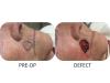
Before
Basal Cell skin cancer above lip Mohs surgery
After
Basal Cell skin cancer above lip Mohs surgery
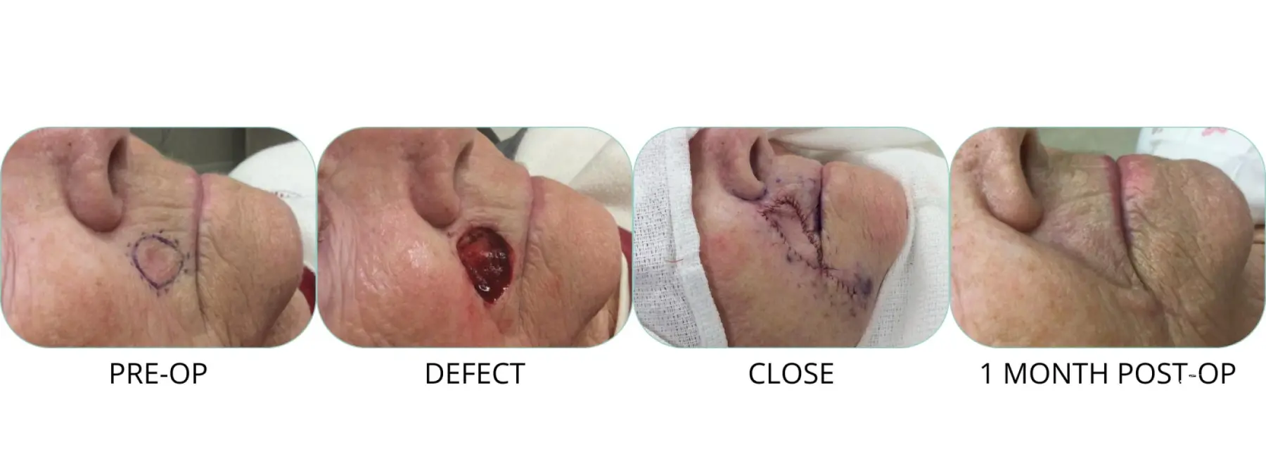
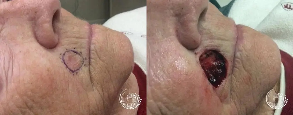
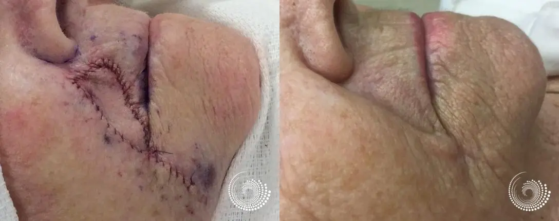
This is an 89-year-old patient was referred for a biopsied skin cancer, a basal cell carcinoma. It was scaly, itchy, and not healing properly.
Pre-op Size: 1.6 cm x 1.4 cm Tissue conservation is critical in areas around the lips.
Removal of the patient's tumor is complicated by the following clinical features: poorly defined clinical tumor borders, large size, clinical area critical for tissue conservation, and located in an area of high recurrence.
Dr. Henry recommended Mohs surgery as the most appropriate treatment for this cancer compared to other treatments. Dr. Henry discussed alternative treatments to Mohs surgery and specifically discussed the risks and benefits of curettage, excision with permanent sections, radiation therapy, and topical chemotherapeutic agents. While these remedies might be effective in some cancers, they have a higher potential of leaving cancerous cells behind, making it necessary for the lesions to be removed again. In fact, these other treatment modalities have a 10 to 15 times higher rate of recurrence when compared to Mohs surgery. The rationale for Mohs was explained to the patient and chosen as the plan to remove this tumor.
Mohs micrographic surgery is a skin cancer treatment that removes all of the cancer, minimizes the risk of recurrence, and leaves as little scarring as possible. Mohs is the most effective treatment for most types of cancer to date with a cure rate of up to 99% for skin cancer. By removing the smallest amount of healthy tissue, it also offers superior cosmetic results. Fellowship-trained American College of Mohs Surgery surgeons, like Dr. Henry, have extensive training in reconstructive surgery and are generally able to perform the reconstructive surgery immediately after microscopic analysis confirms the cancer has been completely removed.
Dr. Henry's compassion and talent are complemented by his elite training as a Fellowship-trained Mohs surgeon, the gold standard for the removal of skin cancers, with minimal healthy tissue impacted, and reconstruction following surgery. There are only 1,500 world-wide Fellowship-trained Mohs surgeons worldwide. Dr. Henry is the most experienced Fellowship-trained Mohs surgeon in this region, having completed over 16,000 Mohs surgeries to date.
MOHS SURGERY STAGE(S):
STAGE 1: The area was prepped, and a rim of normal appearing skin was marked circumferentially around the lesion. The area was infiltrated with local anesthesia. The tumor was first debulked with a curette to remove clinically apparent tumor, tumor visible to the naked eye.
Next an incision was made following the Mohs approach through the skin. The tissue was removed and oriented for further processing. Hemostasis was achieved with electrocautery. The specimen was oriented, mapped and divided into 2 blocks. Frozen section analysis showed: No residual tumor existed.
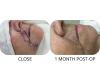
Final Defect Size: 2.5 cm x 1.9 cm
Depth of Final Defect: muscle
For more surgeries, please visit Mohs Surgery Book
- Age: Between 76 and 85 years old
- Post-op Timeline: 1 month
- Technique: Mohs Surgery to Remove Skin Cancer